Equine Complex Vertebral Malformation: Degrees
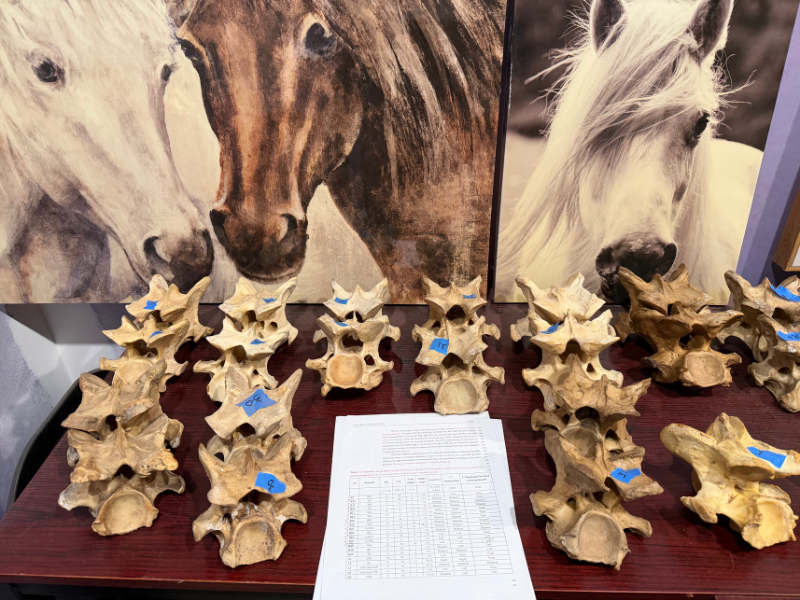
A grading scale for Equine Complex Vertebral Malformation (ECVM) was first described by May-Davis et al. (2023), using postmortem specimens. This system evaluates the degree of absence of the caudal ventral lamina of the sixth cervical vertebra (C6) on a scale from 1 to 4:
- Grade 1: Approximately 25% of the ventral lamina is absent
- Grade 2: Approximately 50% absent
- Grade 3: Approximately 75% absent
- Grade 4: Complete (100%) absence
The Importance of Grading
Grading is an important step in classifying ECVM horses, as evidence suggests that the severity of the absence matters. In these cases, the greater the absence of the caudal (and sometimes cranial) ventral lamina, the higher the incidence of additional changes being present. These can include transposition to the seventh cervical vertebra (C7), which is primarily observed in C6 grade 4s and is associated with a higher likelihood of rib abnormalities and more significant soft-tissue changes, especially in the longus coli.
Clinical and Anatomical Severity
Clinical severity also appears to correlate with anatomical severity. At Rexos Inc., approximately 88% of horses undergoing necropsy due to severe clinical signs and the inability to be safely managed were diagnosed as C6 Grade 4 patients. These horses were classified by radiographic protocols established by De Clue et. al. (2025) and Rexos Inc. This radiographic grading system enabled pre-mortem assessment of each horse after all other differentials were eliminated.
These findings highlight that ECVM cases are not uniform, and further research is needed to better understand the differences among individual horses.
It’s More Than Bone
While the bony changes of ECVM are the most visible and easiest to identify (particularly with imaging), this condition involves more than just bone. Skeletal abnormalities often influence surrounding soft tissues, though not every horse will have the same degree or type of soft-tissue involvement.
Longus Colli
The longus colli is responsible for stabilization and flexion of the cervicothoracic junction. It consists of multiple segments extending from C1 to approximately T5–T6.
The most commonly affected portion is the deep thoracic bundle, which has a strong tendon that normally inserts on the caudal ventral lamina of C6. When this portion of bone is absent, the tendon may be weakened, redirected, or absent. These changes may contribute to instability within the lower cervical spine.
In addition, the longus colli contains a high density of proprioceptive nerve fibers. Changes to this muscle may therefore impact not only biomechanics, but also long-term stability and proprioception.
Scalene Muscles and Brachial Plexus
The dorsal and ventral scalene muscles responsible for lateral flexion of the neck and expansion of the first rib. In some ECVM cases, the dorsal and ventral scalene muscles can have abnormal branching (some bifurcate) and the normal path of the brachial plexus (between the two scalene muscles) deviates through these bifurcations. These variations may contribute to irritation or entrapment of the brachial plexus as a whole or any of the nerves involved in the deviation (CN–6, 7,8 and TN–1,2).
Trachea
The trachea may also be malformed in some horses with ECVM. At this time, the clinical significance of this finding is unknown.
Vascular Changes
In 68% of C7 transpositions, a replication of the foramen transversarium is evident. In these cases, the normal pathway of the cervical vertebral artery from the subclavian artery deviates and enters the cervical column through C7 rather than C6.
This altered path might increase the likelihood of arterial compression during lateral bending of the neck, especially when considering the vertebral artery’s close proximity to the articulation of the 1st sternal rib. In human medicine, similar compression has been associated with serious outcomes such as strokes, though this has not been clearly established in horses.
These vascular differences are also important in surgical planning. Procedures such as foraminotomies may be complicated by abnormal vessel positioning, increasing the risk of bleeding or other complications.
Summary
ECVM is not solely a bone condition. While skeletal changes are the most obvious, associated soft-tissue, neurological, and vascular adaptations play an important role in how each horse is affected.
As with clinical presentation, there is variability between horses. Understanding both the extent of bony changes and the potential for soft-tissue involvement is key to developing a more complete picture of each case.
Read MoreOur Research Paper
